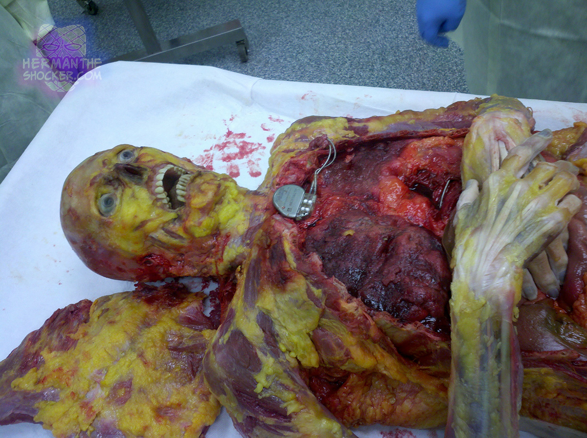
This cadaver is being dissected by medical students. A pacemaker can be seen on the chest. The modern cardiac pacemakers have a smooth external case, with the manufacturer’s data and serial numbers. Leads emerge from one point on the device and pass onwards to the heart. At autopsy, most cases have the generator unit (box) cut free from the wires to allow easy extraction. If the device may have not worked adequately, the entire unit with wires and electrodes needs to be retrieved intact and sent for specialized analysis.

Fig.1 A pacemaker on a cadaver being dissected.

Latest posts

Incomplete skeletonization of a forearm due to post-mortem animal scavenging by a domestic German Shepherd. Skeletonization (synonym: skeletalization)…

USA. A case of simple asphyxia combined with external airway obstruction by a plastic bag, in a suicidal…

This photo depicts a standard “execution type” drug hit. The victim’s hands were secured behind his back. A…

The individual shown in the image committed suicide by shotgun. The image is taken during autopsy. The head…
